Facilities & Scanners
 |
CFMRI - W.M. Keck BuildingThe CFMRI occupies the W.M. Keck Building, an approximately 7,000 sq. ft. building on the main UCSD campus in the School of Medicine adjacent to the Biomedical Science Building. In addition to four magnet bays the facility contains:
|
|
|

|
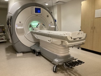
3.0T Scanner (3T West)– Siemens MAGNETOM Cima.X (2025)Installed in Summer 2025, the CFMRI houses a state-of-the-art research dedicated Siemens MAGNETOM Cima.X 3.0T whole-body imaging system for studies in humans and larger animals. The Cima.X system is equipped with:
RF Coils: Available RF coils for the Siemens Cima.X 3T MRI scanner include the Head/Neck 20ch tiltable with CoilShim, Head 32ch, BioMatrix Spine 72ch with Respiratory Sensor, BioMatrix Body 18ch with Beat Sensor (Cardiac triggering without the need for ECG), Body Coil (Standard and Long) 18ch, Shoulder Coil Kit (Small and Large) 16ch, Hand/Wrist 16ch, Knee TX/RX 15ch, Foot/Ankle 16ch, Flex Coil Large & Small 4ch. |
|
|

|
3.0T Scanner (3T East)– Siemens MAGNETOM Prisma (2021)As of 2021, the CFMRI houses a state-of-the-art research dedicated Siemens MAGNETOM Prisma 3.0T whole-body imaging system for studies in humans and larger animals. The Prisma platform is equipped with:
RF Coils: Available RF coils for the Siemens Prisma 3T MRI scanner include Head/Neck 20ch, Head 32ch, Head/Neck 64ch, Body Coil (Standard and Long) 18ch, Spine 32ch Shoulder Coil Kit (Small and Large) 16ch, Hand/Wrist 16ch, Knee TX/RX 15ch, Foot/Ankle 16ch, Flex Coil Large & Small 4ch. Prisma-Available Coils |
|
|
|
3.0T Scanner (3T Keck)– GE MR 750The CFMRI currently houses a state-of-the-art research dedicated General Electric (GE) Discovery MR750 3.0T whole-body imaging system for studies in humans and larger animals. The Discovery MR750 platform is equipped with:
RF Coils: Available RF coils for the 3T MR750 system include the integrated body coil, a Nova Medical 32ch head coil, Invivo 8-channel head coil, GE flexible surface coils, 32ch Body Coil, 16ch Head/Neck Spine Coil, HD 8ch Foot/Ankle Coil, 8ch Wrist Array, HD Shoulder Array, T/R Knee Array,16ch GEM Flex Coil, and an 8ch SENSE Cardiac Coil. |
|
|

|
7.0T Scanner – Bruker - NEO ConsoleThe 7T (20 cm bore) small animal imaging system provides high field strength and gradient performance for high resolution studies of small animals and tissue samples. The main system components are a Bruker 7T magnet with Avance II hardware. We support many imaging applications including:
Radiofrequency coils for multiple applications are available including rat and mouse brain surface coils and three quadrature volume transmit/receive coils of various diameters. We are equipped for both proton and phosporous spectroscopy applications including a phosphorous surface coil and volume resonator and programmable decoupling. Two gradient sets are available for different applications, a 12 cm gradient with 660 mT/m strength and 4570 T/m/s slew rate and a higher performance 6 cm gradient with 1000 mT/m strength and 11250 T/m/s slew rate. Cardiac and respiratory monitoring systems are available for gated acquisition. Temperature monitoring is available for feedback controlled heating. A support cradle with an isoflurane delivery and scavenging system is available. |
|
|

|
Mock ScannerThe CFMRI also houses a Mock Scanner which simulates the look, sound and feel of a real MRI scanner. The mock scanner allows research subjects to experience a simulated MRI scan prior to undergoing the actual MRI scan session, thus increasing the successful rate of data acquisition and the quality of fMRI data. Investigators who have approved or pending research projects at CFMRI may request access to the Mock Scanner. |
|
|

|
Examination Rooms/Waiting AreaThe CFMRI has three Examination Rooms that can be reserved for research participant consultations in parallel with booked scan time on the Center scanners. There is also adequate seating accommodation for research participants in the central waiting area adjacent to the 3.0T scanners.
|
|
|